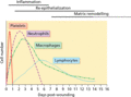
Figure 10.1
Wound inflammatory cells during the first 2 weeks of wound healing showing the stages of acute wound healing and the inflammatory infiltrate present i...
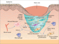
Figure 10.5
Wound contraction to initiate scarring. Macrophage‐derived platelet‐derived growth factor (PDGF), transforming growth factor‐β (TGF‐β) and fibroblast ...
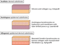
Figure 10.9
Examples of biological skin substitutes.
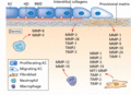
Figure 10.2
Matrix metalloproteinases (MMPs) and tissue inhibitor of matrix metalloproteinases (TIMPs) in wound healing showing the expression and cellular source...
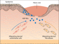
Figure 10.6
Model of how secreted heat shock protein 90α (HSP‐90α) promotes wound repair. Injury to the skin triggers the release of transforming growth factor β ...
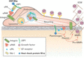
Figure 10.3
Mechanisms involved in matrix degradation during keratinocyte migration. Physiological control of pericellular proteolysis occurs primarily through th...
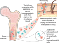
Figure 10.7
Better and faster skin wound healing is achieved by the combined action of AMD3100 and tacrolimus. AMD3100 interferes with the interaction of the CXC ...
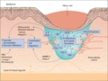
Figure 10.4
Molecular and cellular factors required for angiogenesis in order to promote endothelial cell migration, proliferation and tubule formation. After wou...
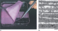
Figure 10.8
Keratinocyte grafting. (a) Sheet of cultured keratinocytes for grafting (b) Ultrastructure of keratinocyte graft. Scale bar 1 μm. (Courtesy of Profes...
