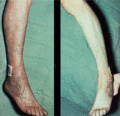
Figure 103.1
Buerger's test showing postural colour change in an ishaemic foot – white when the foot is elevated (right) and red when lowered (left).
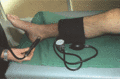
Figure 103.5
Doppler ultrasound to measure the ankle–brachial Doppler pressure index.
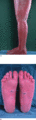
Figure 103.9
Secondary changes in erythromelalgia due to immersion in water to relieve symptoms (a) irritant contact dermatitis, and (b) fissuring. (Courtesy of P...
Figure 103.13
Hereditary haemorrhagic telangiectasia.
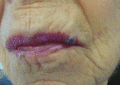
Figure 103.17
Larger venous lakes on upper and lower lips.
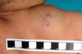
Figure 103.21
Infant with an arteriovenous malformation.
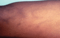
Figure 103.25
Thrombophlebitis migrans.
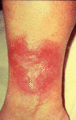
Figure 103.29
Lipodermatosclerosis.
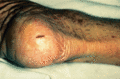
Figure 103.2
Trophic changes including dry skin, cracks, loss of hair and thickened nails (the latter two not shown on figure).
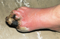
Figure 103.6
Ischaemic toes in Buerger disease.
Figure 103.10
(a) Generalized essential telangiectasia; (b) generalized essential telangiectasia showing blanching with pressure.
Figure 103.14
Spider telangiectases. Classically, when the central arteriole is compressed, the skin blanches and the lesion temporarily vanishes but rapidly return...
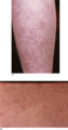
Figure 103.18
Angioma serpiginosum: (a) grouped lesions, and (b) close up appearance.
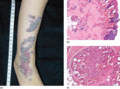
Figure 103.22
Verrucous haemangioma. (a) A linear congenital hyperkeratotic plaque on the lower leg and ankle of a child. (b) Dilated vessels in the superficial der...
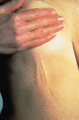
Figure 103.26
Mondor disease.
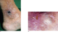
Figure 103.30
(a) Atrophie blanche: white scars with a central ischaemic ulcer and telangiectasia at the edge of the white areas. (b) Venous ulceration with atrophi...
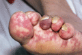
Figure 103.3
Platelet emboli can lodge in the vasculature causing areas of discoloration in the toes and sole of the foot, and can appear ‘vasculitic‐like’.
Figure 103.7
Angiography in thromboangiitis obliterans showing vascular occlusion and corkscrew collaterals.
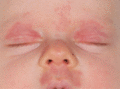
Figure 103.11
Naevus flammeus.
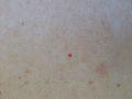
Figure 103.15
Cherry angioma.
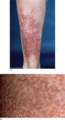
Figure 103.19
Benign essential telangiectasia: (a) arborizing pattern, and (b) close up appearance.
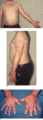
Figure 103.23
Klippel–Trenaunay syndrome: (a) with capillary/venular malformation; (b,c) abnormal veins and soft‐tissue overgrowth.
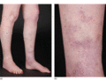
Figure 103.27
(a) Varicose veins in the left leg, and (b) with superficial telangiectasia.
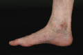
Figure 103.31
Corona phlebectatica paraplantaris.
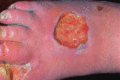
Figure 103.4
Ulceration of the skin at pressure points.
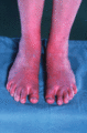
Figure 103.8
Patient during an attack of erythromelalgia.
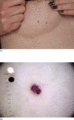
Figure 103.12
(a) Angiokeratoma; (b) dermoscopic view of angiokeratoma.
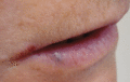
Figure 103.16
Small venous lake.
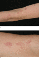
Figure 103.20
(a) Unilateral naevoid telangiectasia, and (b) showing grouped lesions in close up.
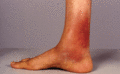
Figure 103.24
Superficial venous thrombosis.
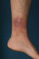
Figure 103.28
Venous eczema.
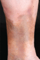
Figure 103.32
Hyperpigmentation secondary to venous insufficiency.
