Figure 109.1
Anatomy of the normal eye. (a) The external appearance of the right eye. (b) Cross‐section of the human eye.

Figure 109.5
Allergic blepharoconjunctivitis due to topical medication toxicity. (a) Allergic eyelid skin reaction. (b) Follicular conjunctival inflammation accomp...

Figure 109.9
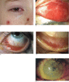
Rarer causes of chronic blepharitis. (a,b) Sebaceous carcinoma of the upper lid. Basal cell carcinomas (BCCs) may also ‘masquerade’ as chronic blephar...

Figure 109.13
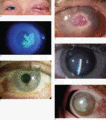
Atopic keratoconjunctivitis (AKC). (a) Normal upper tarsal conjunctiva; the tarsal vessels are clearly visible through the healthy conjunctival epithe...

Figure 109.17
Management algorithm for the management of cicatrizing conjunctivitis. IVIG, intravenous immunoglobulin; MMP, mucous membrane pemphigoid; TNF, tumour ...

Figure 109.21
Molluscum contagiosum at the medial aspect of the upper lid margin with an associated follicular conjunctivitis. (Courtesy of Mr J. Dart, Moorfields ...

Figure 109.25
(a) Posterior subcapsular cataract typically induced by corticosteroid therapy. (b) Papilloedema (swollen optic disc) with accompanying retinal haemor...

Figure 109.29
Cyst of Zeiss. Opaque sebaceous cyst.

Figure 109.33
Basal cell carcinoma (BCC). (a) Ulcerated BCC on the lower lid. (b) Poorly defined BCC at medial canthus. (c) Morphoeic BCC along the lower lid. (Cou...

Figure 109.2
Cross‐section of the upper eyelid.

Figure 109.6
Periorbital oedema due to allergy.

Figure 109.10
Management algorithm for Blepharitis. MGD, meibomian gland dysfunction; FML, fluoromethalone.

Figure 109.14
Atopic blepharoconjunctivitis (ABC).

Figure 109.18
Systemic immunosuppression stepladder for ocular mucous membrane pemphigoid (MMP). Immunosuppression can be carried out using a ‘stepladder’ approach ...

Figure 109.22
Herpes simplex virus. (a) Primary herpes simplex blepharoconjunctivitis in a child. (b) Dendritic epithelial keratitis. (c) Stromal herpetic keratitis...

Figure 109.26
Xanthelasma. There are two lesions on the medial eyelid and one lesion on the temporal eyelid. The cornea shows arcus senilis, which is also associate...

Figure 109.30
(a, b) Naevus of Ota. In this case the pigmentation is confined to the eye, with no cutaneous involvement. The yellow staining of the eyelid is due to...

Figure 109.34
Squamous cell carcinoma (SCC). Infiltrating ulcerated SCC on the lower lid. (Courtesy of Mr N. Joshi, Chelsea and Westminster Hospital/Medical Illust...

Figure 109.3
The lacrimal apparatus of the right eye.

Figure 109.7
Staphylococcal blepharitis. (a) Fibrinous ‘collarettes’ lifting away from the skin as the lashes grow. (b) Fibrinous scales on the anterior lid margin...

Figure 109.11
Seasonal and perennial allergic conjunctivitis (SAC and PAC) (a) Eyelid oedema and redness (b) Upper tarsal papillary inflammation (c) Lower tarsal pa...

Figure 109.15
Management algorithm for allergic eye disease. FML, flouromethalone; HSV, herpes simplex virus; SCG, sodium cromoglycate.

Figure 109.19
Ocular disease in Stevens–Johnson syndrome. (a) Acute conjunctivitis with mucus discharge occurring concurrently with typical erythema multiforme skin...

Figure 109.23
Herpes zoster. (a) Herpes zoster ophthalmicus. (b) Dendritiform keratopathy in herpes. (c) Stromal keratitis (opacity in the central cornea) and uveit...

Figure 109.27
Juvenile xanthogranuloma.

Figure 109.31
Capillary haemangioma. Enlarging lesion on the right upper lid, starting to occlude vision. (Courtesy of Mr N. Joshi, Chelsea and Westminster Hospita...

Figure 109.35
Malignant melanoma. Irregularly pigmented lesion on the lower lid. (Courtesy of Mr N. Joshi, Chelsea and Westminster Hospital/Medical Illustration UK...

Figure 109.4
Madarosis due to staphylococcal blepharitis. (Courtesy of Mr J. Dart, Moorfields Eye Hospital, London, UK.)

Figure 109.8
Ocular rosacea. (a) Scales on the anterior lid margin, meibomitis with posterior migration of the orifices associated with loss of the normal posterio...

Figure 109.12
Vernal keratoconjunctivitis (VKC). (a) ‘Giant’ upper tarsal cobblestone papillae with mucous exudate in palpebral VKC. (b) Typical pale limbal papilla...

Figure 109.16
Ocular signs of mucous membrane pemphigoid (MMP). (a) Inferior fornix shortening and subconjunctival scarring. (b) Conjunctival symblepharon tethering...

Figure 109.20
Wart on the eyelid.

Figure 109.24
(a) External hordeolum (stye). (b) Internal hordeolum (infected chalazion) which has spontaneously discharged anteriorly through the skin, which is no...

Figure 109.28
Cyst of Moll. Small translucent cyst on the anterior lid margin. (Courtesy of Mr N. Joshi, Chelsea and Westminster Hospital/Medical Illustration UK, ...

Figure 109.32
Keratoacanthoma. Keratin‐filled crater on the lid margin. (Courtesy of Mr N. Joshi, Chelsea and Westminster Hospital/Medical Illustration UK, London,...

Figure 109.36
Sebaceous gland carcinoma. Infiltrating lesion on the upper lid. (Courtesy of Mr N. Joshi, Chelsea and Westminster Hospital/Medical Illustration UK, ...

