Figure 123.1
Histologically proven psoriasis appearing in a split‐skin donor site. (Courtesy of Southmead Hospital, Bristol, UK.)
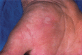
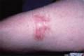
Figure 123.5
Friction blister on the palm mimicking a target lesion of erythema multiforme. This patient had generalized pruritus caused by biliary cirrhosis, and ...
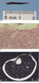
Figure 123.9
Foreign‐body reaction caused by a deeply embedded thorn. The patient presented with a chronic leg ulcer. (a) Thorn that eventually emerged spontaneous...
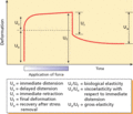
Figure 123.2
Deformation of skin by an applied force showing how elastic and viscoelastic properties can be deduced from ratios of measurements.
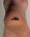
Figure 123.6
Black heel showing stippled pigmentation within the stratum corneum.
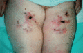
Figure 123.10
Pentazocine ulcers.
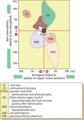
Figure 123.3
Rheological properties of some conditions affecting the dermis. Each area represents data from several patients. (From Piérard et al . [ ]. Reproduc...
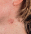
Figure 123.7
‘Fiddler's neck’. Lichenification and cysts on the neck of a violinist. (Courtesy of Dr R. D. G. Peachey, Bristol Royal Infirmary, Bristol, UK.)
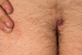
Figure 123.11
Pilonidal sinus.
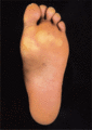
Figure 123.4
Calluses of the forefoot.
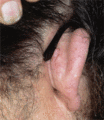
Figure 123.8
Spectacle‐frame acanthoma showing a soft plaque, which may mimic basal cell carcinoma, caused by pressure and friction from the spectacle frame.
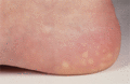
Figure 123.12
Piezogenic pedal papules.
