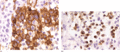
Figure 136.1
Langerhans cell histiocytosis (LCH) lesion in the skin showing (a) characteristic CD1a‐positive staining of LCH cells and (b) skin LCH cells showing c...
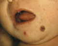
Figure 136.5
Congential self‐healing reticulohistiocytosis (Hashimoto–Pritzker disease) showing ulcerated nodules in an infant.
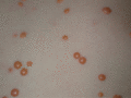
Figure 136.9
Juvenile xanthogranuloma showing multiple, disseminated, yellow papules.
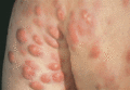
Figure 136.13
Progressive nodular histiocytosis in a 48‐year‐old man with nodular lesions in the posterior axillary fold. (Courtesy of Professor J. M. Naeyaert, Un...
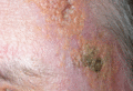
Figure 136.17
Necrobiotic xanthogranuloma showing a reddish yellow, infiltrative plaque with evidence of necrosis. (Courtesy of Dr S. Walsh, Sunnybrook Hospital, T...
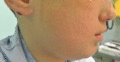
Figure 136.21
Rosai–Dorfman disease showing multiple nodules/tumours with superficial crusting and old scars on the nose.
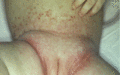
Figure 136.2
Langerhans cell histiocytosis showing a polymorphic eruption of papules, vesicles, crusts and telangiectasias affecting the nappy area, including the ...
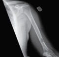
Figure 136.6
Radiograph of a humerus showing a typical punched‐out lytic lesion of bone Langerhans cell histiocytosis.
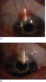
Figure 136.10
Juvenile xanthogranuloma of the iris. (a) Right eye, 4 November 2001. (b) Right eye, July 2003. (Courtesy of Dr J. Donadieu, Trousseau Hospital, Pari...
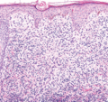
Figure 136.14
Xanthoma disseminatum: similar histological picture to juvenile xanthogranuloma consisting of dermal infiltrate of small spindled histiocytes (H&E). ...
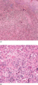
Figure 136.18
Reticulohistiocytoma/multicentric reticulohistiocytosis. (a) The dermis contains an infiltrate of large, eosinophilic, cytoplasm‐rich histiocytes with...
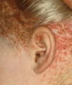
Figure 136.3
Langerhans cell histiocytosis showing yellow crusted papules in a seborrhoeic dermatitis distribution in an older child.
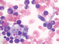
Figure 136.7
Haemophagocytosis in haemophagocytic lymphohistiocytosis.
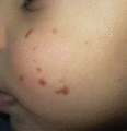
Figure 136.11
Benign cephalic histiocytosis showing multiple, asymptomatic, reddish brown papules on the face of a toddler.
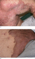
Figure 136.15
(a, b) Xanthoma disseminatum showing large, yellow‐brown plaques affecting the skin folds. (Courtesy of Dr S. Walsh, Sunnybrook Hospital, Toronto, Ca...
Figure 136.19
Multicentric reticulohistiocytosis nodules over the elbows (a) that shrank after an infusion of zoledronic acid (photograph taken 43 days after infusi...
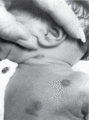
Figure 136.4
Skin‐only Langerhans cell histiocytosis in an infant with spontaneous resolution.
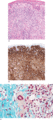
Figure 136.8
Juvenile xanthogranuloma. (a) Small, slightly spindled histiocytes permeate the dermis, splaying collagen fibres. Touton cells are not represented. Ma...
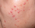
Figure 136.12
Generalized eruptive histiocytosis showing multiple, small, symmetrical, red‐brown papules in an adult. (Courtesy of Dr S. Walsh, Sunnybrook Hospital...
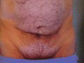
Figure 136.16
Diffuse plane xanthomatosis in a 73‐year‐old man of 7 years’ duration with associated IgG‐κ paraprotein.
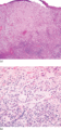
Figure 136.20
Rosai–Dorfman disease. (a) The ‘lymph node‐in‐skin’ appearance is highlighted at a low‐power magnification of 20× (H&E). (b) Rosai–Dorfman histiocytes...
