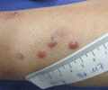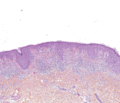
Figure 143.1
Large congenital naevus.

Figure 143.5
(a,b) Superficial spreading melanoma (late presentation).

Figure 143.9
Acral lentiginous melanoma of the sole (late presentation).

Figure 143.13
Amelanotic melanoma. (a) Amelanotic superficial spreading melanoma. (b) Amelanotic nodular.

Figure 143.17
Acral lentiginous melanoma with typical lentiginous pattern.

Figure 143.21
Malignant blue naevus.

Figure 143.25
(a,b) Pigmented melanoma skin metastasis. In a subset of melanoma patients mostly with primary melanoma on the lower extremity, melanoma metastases de...

Figure 143.29
(a,b) Rash secondary to BRAF inhibitor. A patient treated with the selective BRAF inhibitor vemurafenib (after 2 weeks of treatment) has developed...

Figure 143.2
Atypical/dysplastic naevi.

Figure 143.6
(a–c) Nodular melanomas.

Figure 143.10
Acral lentiginous melanoma in the nail area. Early presentation as a longitudinal melanonychia.

Figure 143.14
(a,b) Amelanotic acral lentiginous melanomas.

Figure 143.18
Lentigo maligna melanoma.

Figure 143.22
Breslow depth.

Figure 143.26
Amelanotic melanoma skin metastasis. Amelanotic melanoma skin metastases require a very careful skin examination, as they can be easily missed.

Figure 143.30
Positron emission tomography‐computed tomography (PET‐CT) scans of an advanced BRAF mutated melanoma patient treated with selective BRAF inhibitor v...

Figure 143.3
Atypical/dysplastic naevus syndrome.

Figure 143.7
(a–e) Lentigo maligna melanomas.

Figure 143.11
(a–c) Acral lentiginous melanomas in the nail area. Late presentation.

Figure 143.15
Regressive mealnomas. (a) Partial regression. (b) Quasi‐total regression.

Figure 143.19
Superficial spreading melanoma.

Figure 143.23
(a–c) Sentinel lymph node biopsy allows detection of melanoma micrometastases in regional lymph nodes. Radioactive tracer and dye are injected intrade...

Figure 143.27
Targeting signalling pathways in melanoma. The mitogen‐activated protein kinase (MAPK) pathway is one of the most important signalling pathways in mel...

Figure 143.4
(a–c) Superficial spreading melanoma (early presentation).

Figure 143.8
(a–d) Acral lentiginous melanomas.

Figure 143.12
Mucosal melanomas. (a) Anal; (b) oral.

Figure 143.16
Nodular melanoma.

Figure 143.20
Desmoplastic melanoma.

Figure 143.24
Treatment algorithm of advanced melanoma in a tertiary referral centre. The landscape of systemic treatment options in metastatic melanoma has rapidly...

Figure 143.28
The mitogen‐activated protein kinase (MAPK) pathway. This pathway is a key regulator for gene expression that drives melanocytic gene differentiation ...

