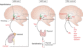
Figure 149.1
Schematic representation of three hypothalamopituitary axes that impact on human skin: key elements of the central hypothalamopituitary–adrenal (HPA) ...
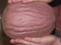
Figure 149.5
Cutis verticis gyrata.
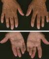
Figure 149.9
Addisonian pigmentation of the palmar creases 37 years after bilateral adrenalectomy for Cushing disease from ACTH‐producing pituitary adenoma (Nelson...
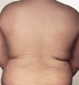
Figure 149.13
Striae due to obesity in a young man.
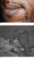
Figure 149.17
Neurosarcoidosis with hypopituitarism presenting as a rash. A diagnosis of cutaneous and pulmonary sarcoidosis was readily established in a 35‐year‐ol...
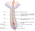
Figure 149.2
The human hair follicle as a (neuro‐) endocrine microcosm. This schematic drawing of a human anagen VI scalp hair follicle indicates the main intrafol...
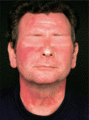
Figure 149.6
Histamine‐evoked ‘geographical’ pattern of flushing due to foregut carcinoid tumour. (Courtesy of Professor M. Greaves, London, UK.)

Figure 149.10
Melasma.

Figure 149.14
Pigmentation of (a) the gingivae and (b) the tongue in a woman who presented with darkening skin due to Addison disease.

Figure 149.3
Acanthosis nigricans, skin tags and striae in a 41‐year‐old obese male with type 2 diabetes.
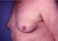
Figure 149.7
Gynaecomastia due to long‐term spironolactone therapy given for hypertension.
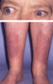
Figure 149.11
A patient with Graves disease with pretibial myxoedema and exophthalmos.

Figure 149.15
Acromegalic macroglossia.
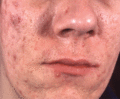
Figure 149.4
Acromegaly: note the coarse features, severe acne and seborrhoea.
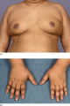
Figure 149.8
An 18‐year‐old male with pituitary Cushing disease followed by hypopituitarism. (a) Obesity and gynaecomastia. (b) Insulin resistance with acanthosis ...
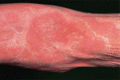
Figure 149.12
Necrolytic migratory erythema. (Courtesy of Dr Kristian Thomsen, Finsen Institute, Copenhagen, Denmark.)
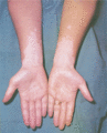
Figure 149.16
Vitiligo.
