Figure 24.1
Approximate per cent depth dose values for 90 kV superficial X‐ray beam.

Figure 24.5
(a) ‘Bathing cap’ distribution showing minimal radiotherapy dose to the eyes, optic nerves and chiasm. (b) Bathing cap showing high radiotherapy dose ...

Figure 24.9
(a) SCC on the back, unsuitable for radiotherapy. (b) SCC on the buttock not suitable for radiotherapy. The sites shown in (a) and (b) tolerate radiot...

Figure 24.13
(a) BCC of the posterior aspect of the ear. (b) The same patient 6 months after electron beam treatment.

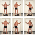
Figure 24.17
(a–f) The modified Stanford technique. Patient positions to optimize a uniform dose to all the skin when using total skin electron beam therapy (TSEBT...

Figure 24.21
Telangiectasia after treatment for a small BCC of the nose.

Figure 24.2
Approximate per cent depth dose curves for 4.5 and 9 MeV electron beams.

Figure 24.6
(a) Homogeneous dose distribution over the skin of the foot, ankle and lower leg using TomoTherapy. (b) Dose distribution for TomoTherapy treatment of...

Figure 24.10
(a) BCC of the lower eyelid. (b) The same patient 6 months after superficial X‐ray treatment.

Figure 24.14
(a) Large BCC pre‐auricular skin. (b) The same patient 6 months after superficial X‐ray treatment.

Figure 24.18
(a) Large BCC of the cheek. (b) The same patient showing acute radiation reaction 6 weeks after superficial X‐ray treatment. (c) The same patient show...

Figure 24.3
Superficial X‐ray machine.

Figure 24.7
Late sequelae after radiation treatment for ringworm – atrophy, pigmentation change, alopecia, telangiectasia and multiple BCCs.

Figure 24.11
(a) BCC of the upper inner canthus. (b) The same patient 5 years after superficial X‐ray treatment.

Figure 24.15
(a) SCC of the lip. (b) The same patient 8 years after higher energy kilovoltage treatment.

Figure 24.19
(a) Large BCC of the jaw. (b) The same patient showing acute radiation reaction 4 weeks after superficial X‐ray treatment. (c) The same patient showin...

Figure 24.4
(a) Lead mask with area for treatment ‘cut‐out’. (b) BCC to be treated in this manner.

Figure 24.8
(a) SCC too large to expect long‐term cure. (b) Satisfactory control at 6 months.

Figure 24.12
(a) BCC of the upper pinna. (b) The same patient 6 weeks after electron beam therapy.

Figure 24.16
(a) Malignant melanoma, pre‐radiotherapy. (b) Malignant melanoma, post‐radiotherapy.

Figure 24.20
(a) Extensive erosive BCC of the ear. (b) The same patient 2 years after superficial X‐ray treatment.

