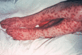
Figure 25.1
Smallpox. Papulovesicular lesions, some with haemorrhagic centres, concentrated on the extremities. (Courtesy of Dr Colin Long, Cardiff and Vale NHS ...
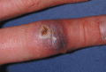
Figure 25.5
Orf. (Courtesy of Addenbrooke's Hospital, Cambridge, UK.)
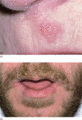
Figure 25.9
Herpes labialis. (a) Typical recurrent lesion on the upper lip. (b) More widespread recurrent lesions following streptococcal pyoderma with lymphangit...
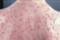
Figure 25.13
Varicella. (Courtesy of York District Hospital, UK.)
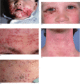
Figure 25.17
Eczema herpeticum. (a) Perioral. (b) Periocular. (c) Forehead. (d) Front of the neck of 20‐year‐old man. (e) Resolving lesions. (Part (d) courtesy of ...
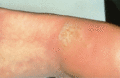
Figure 25.21
Mosaic plantar wart. (Courtesy of Addenbrooke's Hospital, Cambridge, UK.)
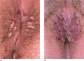
Figure 25.25
(a,b) Perianal warts. (Courtesy of York District Hospital, UK.)
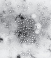
Figure 25.29
Human parvovirus (B19) in the serum of a patient with aplastic crisis. Negatively stained electron micrograph, ×200 000. (Courtesy of Mr T. W. Lee, J...
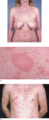
Figure 25.33
Pityriasis rosea: (a) with herald patch on the right of the abdomen, shown in close‐up in (b). (Courtesy of York District Hospital, UK.) (c) With her...
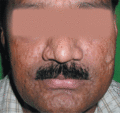
Figure 25.2
Scarring of face following smallpox infection. (Courtesy of Dr S. B. Verma, Baroda Skin Clinic, Baroda, India.)
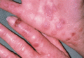
Figure 25.6
Orf with erythema multiforme. The orf lesion on the dorsum of the forefinger has been present for 14 days; the secondary erythema multiforme for 4 day...
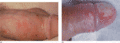
Figure 25.10
Herpes genitalis. (a) Scattered lesions on the penile shaft. (b) Confluent lesions resulting in large erosions. (Courtesy of Addenbrooke's Hospital, ...
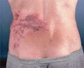
Figure 25.14
Zoster of the trunk. (Courtesy of York District Hospital, UK.)
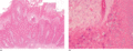
Figure 25.18
Histology of viral wart (a) low power, ×40, showing morphology of wart lesion with papillomatous acanthosis and hyperkeratosis; (b) high power, ×200, ...
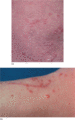
Figure 25.22
Plane warts. (a) Warts on the knee. (b) Warts on the arm with spread into a scratch. (Courtesy of Addenbrooke's Hospital, Cambridge, UK.)
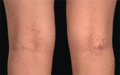
Figure 25.26
Epidermodysplasia verruciformis. Pigmented flat warty lesions in popliteal fossae. (Courtesy of Addenbrooke's Hospital, Cambridge, UK.)
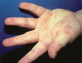
Figure 25.30
Hand, foot and mouth disease. (Courtesy of Addenbrooke's Hospital, Cambridge, UK.)
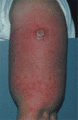
Figure 25.3
Vaccinia: vaccination site with generalized spread. (Courtesy of Addenbrooke's Hospital, Cambridge, UK.)
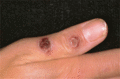
Figure 25.7
Milker's nodule. (Courtesy of Dr J. B. Kurtz, Oxford, UK.)
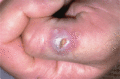
Figure 25.11
Herpes simplex. Inoculation lesion on the thumb of a dermatologist. (Courtesy of Dr A. S. Highet, York District Hospital, UK.)
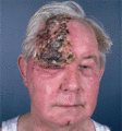
Figure 25.15
Ophthalmic zoster. (Courtesy of York District Hospital, UK.)
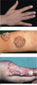
Figure 25.19
Common warts. (a) Hand. (Courtesy of Addenbrooke's Hospital, Cambridge, UK.) (b) Dorsum of the finger, filiform warts. (Courtesy of Dr A. S. Highet, ...
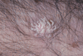
Figure 25.23
Filiform wart on the forearm. (Courtesy of Addenbrooke's Hospital, Cambridge, UK.)
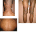
Figure 25.27
Acquired epidermodysplasia verruciformis. Widespread flat hyper‐ and hypopigmented lesions affecting the whole body: (a) neck, (b) back and (c) legs. ...
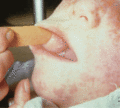
Figure 25.31
Koplik spots on the buccal mucosa in measles. (Courtesy of Dr John Kurtz, Oxford, UK.)
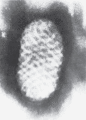
Figure 25.4
Orf virus. Phosphotungstate preparation (×230 000). (Courtesy of Dr J. Nagington, Cambridge, UK.)
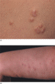
Figure 25.8
Molluscum contagiosum. (a) Typical umbilicated lesions. (b) Depressed scars following infection. (Courtesy of Addenbrooke's Hospital, Cambridge, UK.)...
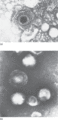
Figure 25.12
Herpesvirus varicellae . Phosphotungstate preparations from vesicle fluid. (a) The dark centre is due to penetration of the capsid by phosphotungstate...
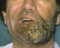
Figure 25.16
Herpes zoster oticus showing unilateral zoster with facial palsy (Ramsay Hunt syndrome).
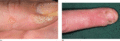
Figure 25.20
(a) Periungual warts. (Courtesy of Addenbrooke's Hospital, Cambridge, UK.) (b) Periungual warts in a nail‐biter. (Courtesy of York District Hospital,...
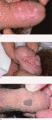
Figure 25.24
Penile warts. (a,b) Classical condylomata acuminata. (c) This pigmented lesion was confirmed histologically to be a viral wart. (Courtesy of York Dis...
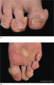
Figure 25.28
Extensive plantar warts in a renal transplant recipient. (a) Toes. (b) Sole. (Courtesy of Addenbrooke's Hospital, Cambridge, UK.)
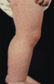
Figure 25.32
Gianotti–Crosti syndrome. Papular eruption on the leg. (Courtesy of Dr N. P. Burrows, Addenbrooke's Hospital, Cambridge, UK.)
