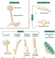
Figure 32.1
Asexual reproduction in fungi. (a) Zygomycota : the most characteristic form of asexual reproduction is the production of sporangia and sporangiospor...
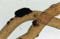
Figure 32.5
Black piedra. Hairs mounted in 30% KOH, bright field. The dark nodules are formed of dematiaceous hyphae cemented together to form a hard mass. (Cour...
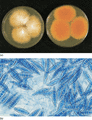
Figure 32.9
Microsporum canis . (a) Colony; the reverse of the colony is shown on the right. (b) Microscopy, bright field. The macroconidia are characteristic, th...
Figure 32.13
Trichophyton interdigitale . (a) Typical colony form; the reverse of the colony is shown on the right. (b) More unusual colony forms. The upper colony...
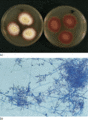
Figure 32.17
Trichophyton tonsurans . (a) Colony; the reverse of the colony is shown on the right. (b) Microscopy, bright field. The microconidia are large and var...
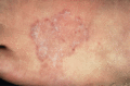
Figure 32.21
Tinea circinata: characteristic ringworm lesions.
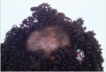
Figure 32.25
Kerion in a patient with Trichophyton tonsurans infection of the scalp.
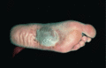
Figure 32.29
Trichophyton interdigitale infection: bullous lesion on the sole.
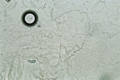
Figure 32.33
Infection by Neoscytalidium species . Skin scales are mounted in 30% KOH, bright field. The hyaline hyphae superficially resemble those of dermatophy...
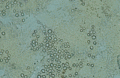
Figure 32.37
Scopulariopsis brevicaulis . Nail clipping mounted in KOH, bright field. The characteristic conidia are relatively thick‐walled, oval or lemon‐shaped ...
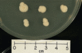
Figure 32.41
Candida albicans colonies showing a white to cream colour on glucose—peptone agar. (Courtesy of the Department of Medical Mycology, St John's Instit...
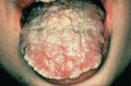
Figure 32.45
Chronic oral candidosis.
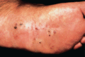
Figure 32.49
Mycetoma caused by Madurella grisea .
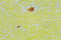
Figure 32.53
Chromoblastomycosis: tissue section. The natural brown pigment of the fungal muriform cells is clearly visible. The cells divide by fission and may fo...
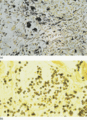
Figure 32.57
Histoplasmosis capsulatum var. capsulatum . (a) Tissue section. The tiny yeasts, stained black with GMS, are largely intracellular. (b) Oil immersio...
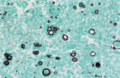
Figure 32.61
Paracoccidioidomycosis. The yeasts, stained black with GMS, are characterized by the numerous peripheral buds produced. (Courtesy of the Department o...
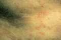
Figure 32.2
Pityriasis versicolor showing typical fine scaling.
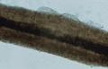
Figure 32.6
White piedra. Hair mounted in KOH, bright field. The gelatinous nodules formed by various Trichosporon species surround the hair. (Courtesy of the ...
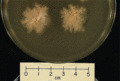
Figure 32.10
Microsporum canis glabrous form colony. (Courtesy of the Department of Medical Mycology, St John's Institute of Dermatology, King's College London, ...
Figure 32.14
Trichophyton rubrum . (a) Downy colony; the reverse of the colony is shown on the right. (b) Downy form microscopy, bright field. Clavate to elongate ...
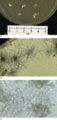
Figure 32.18
Trichophyton verrucosum . (a) Colony. (b) Microscopy at 26°C, bright field. Through the back of the culture dish, the small colonies are characterized...
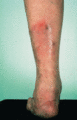
Figure 32.22
Nodular folliculitis caused by Trichophyton rubrum .
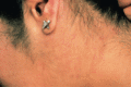
Figure 32.26
Tinea faciei caused by Trichophyton rubrum .
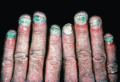
Figure 32.30
Onychomycosis caused by Trichophyton rubrum .
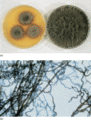
Figure 32.34
Neoscytalidium dimidiatum . (a) Colony forms. The fast‐growing form (right) fills a 90 mm Petri dish in a few days and develops profuse aerial myceliu...
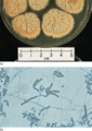
Figure 32.38
Scopulariopsis brevicaulis . (a) Colony. On media free of cycloheximide, the colonies are initially waxy and deeply folded, but the production of coni...
Figure 32.42
Specific identification of Candida albicans can be made by the observation of filaments with thick‐walled terminal vesicles when cultured on a deple...
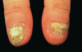
Figure 32.46
Candida onychomycosis in a patient with chronic mucocutaneous candidosis.
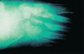
Figure 32.50
Erosive X‐ray changes in a mycetoma.
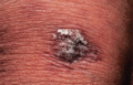
Figure 32.54
Early lesion of chromoblastomycosis.
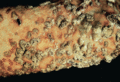
Figure 32.58
Cutaneous blastomycosis. (Courtesy of Dr M. James, Royal Berkshire Hospital, Reading, UK.)
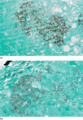
Figure 32.62
Infection by Talaromyces (previously Penicillium ) marneffei . (a) Tissue section. The tiny organisms are largely intracellular and look very simi...
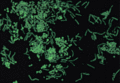
Figure 32.3
Pityriasis versicolor. Skin scales mounted in potassium hydroxide (KOH) and Calcofluor white UV illumination. The hyphae diagnostic of the condition h...
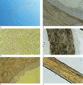
Figure 32.7
Dermatophytosis. (a) Skin scales mounted in 30% KOH, Nomarski illumination. The hyphae are very even in diameter and regularly septate. (b) Skin scale...
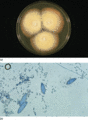
Figure 32.11
Microsporum gypseum . (a) Colony. (b) Microscopy, bright field. The macroconidia are abundant and characteristic, with a symmetrical cigar shape and t...
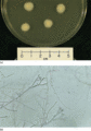
Figure 32.15
Trichophyton schoenleinii . (a) Colony. (b) Microscopy, bright field. Typical antler hyphae may be observed by examining through the back of the cultu...
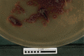
Figure 32.19
Trichophyton violaceum colony. (Courtesy of the Department of Medical Mycology, St John's Institute of Dermatology, King's College London, London, U...
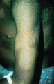
Figure 32.23
Tinea imbricata affecting the upper arm.
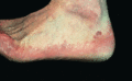
Figure 32.27
Dry‐type Trichophyton rubrum infection.
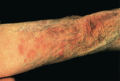
Figure 32.31
Tinea corporis in a patient on systemic corticosteroids.
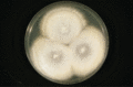
Figure 32.35
Neoscytalidium hyalinum colony. (Courtesy of the Department of Medical Mycology, St John's Institute of Dermatology, King's College London, London, ...
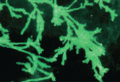
Figure 32.39
Superficial white onychomycosis caused by non‐dermatophyte moulds. Nail clipping mounted in KOH and calcofluor white, UV illumination. Bizarre, frondi...
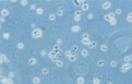
Figure 32.43
A second method for the specific identification of Candida albicans is the observation of germ tubes in serum after incubation at 37°C for 2–4 h. (...
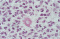
Figure 32.47
Sporotrichosis: tissue section stained with haematoxylin and eosin (H&E). Organisms are usually very scanty but asteroid bodies, representing a foreig...
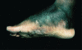
Figure 32.51
Eumycetoma affecting the foot.
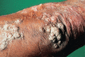
Figure 32.55
Plaque‐type chromoblastomycosis.
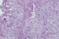
Figure 32.59
North American blastomycosis: tissue section. The large yeasts, stained pink with PAS, are characterized by the broad base of the buds. (Courtesy of ...
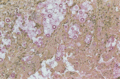
Figure 32.63
Cryptococcosis: tissue section. The mucicarmine stains the capsule specifically. The radiate spiny appearance is caused by shrinkage during processing...
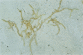
Figure 32.4
Tinea nigra. Skin scales mounted in 30% potassium hydroxide, bright field. The natural brown colour of the septate hyphae is apparent. (Courtesy of t...
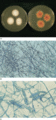
Figure 32.8
Microsporum audouinii . (a) Colony; the reverse of the colony is shown on the right. (b) Microscopy, bright field. In many isolates chlamydoconidia, r...
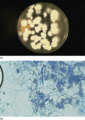
Figure 32.12
Trichophyton mentagrophytes . (a) Colony. (b) Microscopy, bright field. Round microconidia are arranged in clusters; spiral hyphae and thin‐walled, sm...
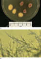
Figure 32.16
Trichophyton rubrum of African origin (formerly called T. soudanense ). (a) Colony forms. A typical apricot yellow and a red isolate are illustrated...
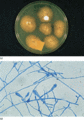
Figure 32.20
Epidermophyton floccosum . (a) Colony. (b) Microscopy, bright field. The clavate macroconidia are characteristic. Microconidia are absent but chlamydo...
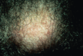
Figure 32.24
Tinea capitis caused by Microsporum canis .
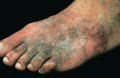
Figure 32.28
Tinea pedis spreading to the dorsum of the foot.
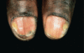
Figure 32.32
Onychomycosis caused by Neoscytalidium dimidiatum : early onycholysis.
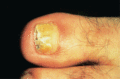
Figure 32.36
Onychomycosis caused by Scopulariopsis brevicaulis .
Figure 32.40
Candidosis. Skin scales mounted in 30% KOH, Nomarski illumination, oil. Budding yeasts and slender filaments are observed. (Courtesy of the Departmen...
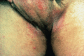
Figure 32.44
Candida infection of the groins
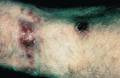
Figure 32.48
Lymphangitic sporotrichosis.
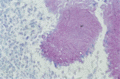
Figure 32.52
Mycetoma: tissue section. Distinct hyphal filaments, stained pink with periodic acid–Schiff (PAS), are clearly visible in a pale‐grained eumycetoma. ...
Figure 32.56
Subcutaneous Basidiobolus infection tissue section. Wide, aseptate hyphae stained black with Grocott methenamine silver (GMS) are characteristic of in...
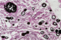
Figure 32.60
Coccidioidomycosis tissue section. Spherules of various sizes are stained black with GMS. In the top left‐hand part of the slide, freshly released end...
