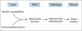
Figure 41.1
Schematic of the pathophysiology of atopic eczema.
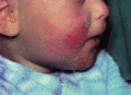
Figure 41.5
Atopic eczema: infantile phase.
Figure 41.9
Atopic ’dirty neck’; reticulate pigmentation on the neck of a patient with longstanding atopic eczema.
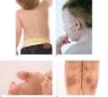
Figure 41.13
(a) Discoid eczema lesions in an atopic child. (b) Discoid lesions on the face. Saliva is a common irritant in young children. (c) Discoid lesion aggr...

Figure 41.17
(a) Follicular lichenification on the surface. (b) Atopic hand eczema.
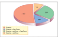
Figure 41.21
The incidence of atopic respiratory symptoms in 1200 patients with atopic eczema. (From Rajka 1960–1 [ ]. Reproduced with permission from Medical Jou...
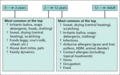
Figure 41.25
Spectrum of trigger factors at different ages. (Adapted from Katayama et al . 2011 [ ]. Reproduced with permission from the Japanese Society of Alle...
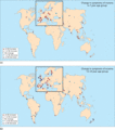
Figure 41.2
(a,b) Worldwide change in atopic eczema symptom prevalence between International Study of Asthma and Allergies in Childhood (ISAAC) Phases 1 and 3. SE...
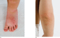
Figure 41.6
(a) Dermatitis causing hypopigmentation. (b) Extensor dermatitis in an infant.
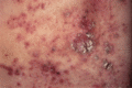
Figure 41.10
Atopic eczema: erythema, papules, excoriations, crusting and secondary infection but, in this case, little lichenification.
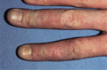
Figure 41.14
Atopic eczema of the fingers of a child.
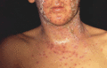
Figure 41.18
Kaposi varicelliform eruption: eczema herpeticum.
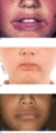
Figure 41.22
(a) Lip‐lick cheilitis. (b) Lip‐lick dermatitis with mild impetiginization. (c) Lip‐lick dermatitis with hyperpigmentation.
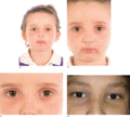
Figure 41.26
(a) Airborne facial eczema with lip‐lick cheilitis. (b) Airborne facial dermatitis with lip‐lick cheilitis. (c) Periorbital dermatitis in the older ch...
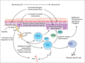
Figure 41.3
Pathophysiology of atopic eczema (AE). LN, lymph node; TSLP, thymic stromal lymphopoietin.
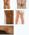
Figure 41.7
(a) Lichenification, crusting and excoriation in the popliteal fossae. (b) Postinflammatory pigmentation. (c) Flexural dermatitis causing hypopigmenta...
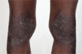
Figure 41.11
Marked lichenification on the knees of an African child. The popliteal fossae were spared.
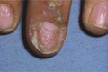
Figure 41.15
Nail involvement in atopic eczema in childhood.
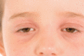
Figure 41.19
Periorbital dermatitis with Dennie–Morgan fold.
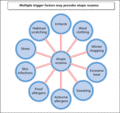
Figure 41.23
Trigger factors for atopic eczema.
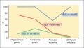
Figure 41.4
Filaggrin (FLG). A representation of the clinical features identified in FLG homozygote or compound hetrerozygote mutations (FLG –/–) versus heteroz...
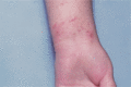
Figure 41.8
Flexural atopic eczema of the wrist in a child.
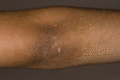
Figure 41.12
Persistent lichenification in an extensor distribution.
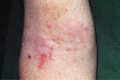
Figure 41.16
Adult flexural dermatitis.
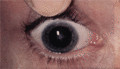
Figure 41.20
Atopic cataract.
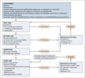
Figure 41.24
Atopic eczema treatment algorithm. TCI, topical calcineurin inhibitors; TCS, topical corticosteroids.
