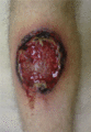
Figure 49.1
Classical pyoderma gangrenosum.
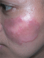
Figure 49.5
Sweet syndrome. The face is often affected.
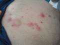
Figure 49.9
BADAS showing crops of large pustules on an erythematous purpuric base on the trunk.
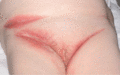
Figure 49.13
Subcorneal pustular dermatosis, showing flexural predistribution notably in the groins.
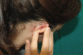
Figure 49.17
Amicrobial pustulosis of the skin folds. Erosions and crusts extensively involving the retro‐auricular region. (Courtesy of Professor A. V. Marzano, ...
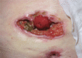
Figure 49.2
Parastomal pyoderma gangrenosum.
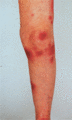
Figure 49.6
Sweet syndrome. Multiple large erythematous lesions on the leg.
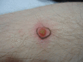
Figure 49.10
A typical initial lesion in BADAS showing a large deep pustule on an erythematous base.
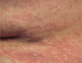
Figure 49.14
Typical appearance of pustules in subcorneal pustular dermatosis.
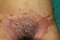
Figure 49.18
Amicrobial pustulosis of the inguinal folds. Erosive and exudating lesions, partially covered by crusts, symmetrically involving the inguinal folds an...
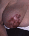
Figure 49.3
Superficial granulomatous pyoderma gangrenosum.
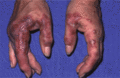
Figure 49.7
Neutrophilic dermatosis of the dorsal hands.
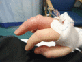
Figure 49.11
Acute inflammatory distal arthritis with tenderness and swelling in BADAS.
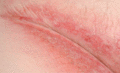
Figure 49.15
Subcorneal pustular dermatosis. Pustules may rupture and be inconspicuous in the skin folds.
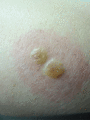
Figure 49.4
Sweet syndrome. Pseudovesicles may occur within the inflammatory plaques.
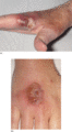
Figure 49.8
(a,b) Bullous variants of neutrophilic dermatosis of the dorsal hands.
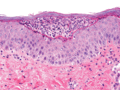
Figure 49.12
Subcorneal pustular dermatosis. Histology showing subcorneal neutrophils. Pustules sit on the surface of the epidermis without spongiosis or acantholy...
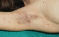
Figure 49.16
Sterile pustules, erosions and crusts on the axillary fold of a patient with amicrobial pustulosis of the skin folds. (Courtesy of Professor A. V. Ma...
