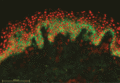
Figure 50.1
Direct immunofluorescence of pemphigus vulgaris. Antibody is deposited around the cell membrane of epidermal keratinocytes.
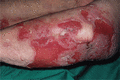
Figure 50.5
Pemphigus vulgaris. Because bullae occur within the epidermis they are fragile and frequently break down to leave widespread erosions. (Courtesy of D...
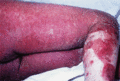
Figure 50.9
Pemphigus foliaceus. Occasionally, pemphigus foliaceus becomes widespread and can result in erythroderma.
Figure 50.13
Serological screening for autoantibodies in pemphigoid diseases: indirect immunofluorescence microscopy on 1 M NaCl‐split human skin. Autoantibodies a...
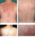
Figure 50.17
Clinical variants of bullous pemphigoid. Eczematous lesions with some erosions and crusts (a,b), prurigo‐like variant with multiple excoriated papules...
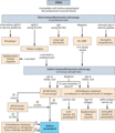
Figure 50.21
Diagnostic pathway for bullous pemphigoid. The diagnostic gold standard is still direct immunofluorescence (IF) microscopy of a perilesional biopsy. A...
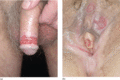
Figure 50.25
Genital involvement in mucous membrane pemphigoid: (a) male, and (b) female.
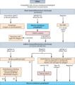
Figure 50.29
Diagnostic pathway for mucous membrane pemphigoid. (1) Overlap with linear IgA disease (with exclusive IgA reactivity) and EBA (with reactivity agains...
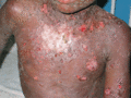
Figure 50.33
Linear IgA disease. Erosions and tense blisters on the trunk in a Ugandan child. Lesions were also present on the face.
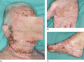
Figure 50.37
Anti‐p200 pemphigoid. Erosions, haemorrhagic crusts and tense blisters on the face (a), palm (b) and foot (c).
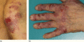
Figure 50.41
Epidermolysis bullosa acquisita, mechanobullous variant. Erythema, erosions and crusts on the left knee (a) and erythematous plaques, atrophic scars, ...
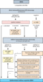
Figure 50.45
Diagnostic pathway for epidermolysis bullosa acquisita (EBA). (1) commercially available; (2) depending of availability; positivity in any of the four...
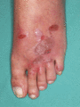
Figure 50.49
Lichen planus pemphigoides. Erosions, erythema, partly ruptured and subsequently desiccated blisters, and a tense vesicle on the left foot. In additio...
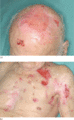
Figure 50.53
Brunsting–Perry pemphigoid. Erosions on the scalp (a), chest and upper left arm as well as atrophic scars (b) in a 95‐year‐old man. Linear staining of...
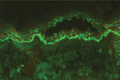
Figure 50.2
Direct immunofluorescence of paraneoplastic pemphigus. Intercellular antibody deposition is seen as in pemphigus vulgaris but in addition there is lab...
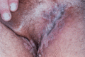
Figure 50.6
Pemphigus vegetans. Vegetating lesions typically occur in the flexures, often without evident blistering. (Courtesy of Dr R.J. Pye, Addenbrooke's Hos...
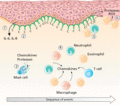
Figure 50.10
Sequence of events leading to blister formation in bullous pemphigoid. Binding of autoantibodies (green) against BP180 (orange) initiates Fc‐independe...
Figure 50.14
Serological screening for autoantibodies in pemphigoid diseases: indirect immunofluorescence microscopy using a BIOCHIP Mosaic™. Ten incubation fields...
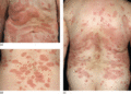
Figure 50.18
Clinical variants of bullous pemphigoid. Urticarial and erythematous plaques (a–c) accompanied by erosions and excoriations (c).
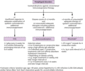
Figure 50.22
Indications and protocols for the treatment of refractory pemphigoid diseases with intravenous immunoglobulin (IVIG), immunoadsorption and rituximab. ...
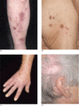
Figure 50.26
Skin lesions in mucous membrane pemphigoid. Brownish erythema, erosions, and some crusts on the right shin (a). Erosions, ulcerations and some atrophi...
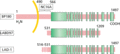
Figure 50.30
Schematic diagram of BP180 (type VII collagen) and its cell‐derived fragments recognized by linear IgA disease (LAD) sera. Most LAD sera contain IgA r...
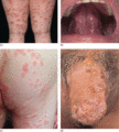
Figure 50.34
Linear IgA disease. Tense blisters in an annular pattern on the thighs (a), erosions on the tongue (b), erythema and blisters on the right gluteal reg...
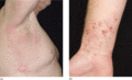
Figure 50.38
Anti‐p200 pemphigoid. Erythematous partly excoriated papules and erythema on the right axilla (a) and vesicles and erythematosus papules on the right ...
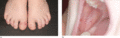
Figure 50.42
Childhood epidermolysis bullosa acquisita, mechanobullous variant. Erythema, erosions and tense blisters on the trauma‐prone dorsal aspects of the toe...
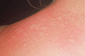
Figure 50.46
Bullous systemic lupus erythematosus. Tense blisters on erythematosus skin.
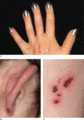
Figure 50.50
Cicatricial pemphigoid. A 36‐year‐old woman with tense vesicles between the thumb and ring finger and several atrophic papules and plaques on the dors...
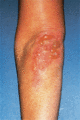
Figure 50.54
Dermatitis herpetiformis. Intact tense bullae on the elbow. (Courtesy of Dr R.J. Pye, Addenbrooke's Hospital, Cambridge, UK.)
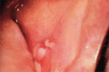
Figure 50.3
Pemphigus vulgaris. Mucosal erosions are an early sign in pemphigus vulgaris, often preceding the cutaneous changes. (Courtesy of Dr R.J. Pye, Addenb...
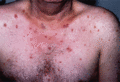
Figure 50.7
Pemphigus foliaceus. There are superficial erosions, frequently without obvious bullae.
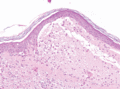
Figure 50.11
Histopathology of bullous pemphigoid. Lesional skin biopsy with subepidermal splitting and a dense inflammatory infiltrate of eosinophils and neutroph...
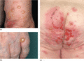
Figure 50.15
Classical bullous pemphigoid. Tense blisters and erosions on the arm (a), hand (b) and gluteal region (c). Blisters may arise on erythematous (a,c) or...
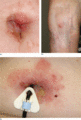
Figure 50.19
Localized bullous pemphigoid. Tense blisters and erosions limited to the umbilical area (a). Single tense blister at the site of major surgery (b). Ec...
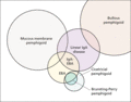
Figure 50.23
Diagnostic overlap between mucous membrane pemphigoid, linear IgA disease, epidermolysis bullosa acquisita (EBA), cicatricial pemphigoid and bullous p...
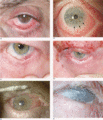
Figure 50.27
Ocular disease in mucous membrane pemphigoid. Conjunctival hyperaemia, inferior fornix shortening and loss of the plica in early disease (Foster II, M...
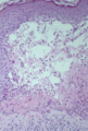
Figure 50.31
Lesional histopathology of linear IgA disease. H&E staining shows subepidermal splitting with a dense inflammatory infiltrate in the blister cavity an...
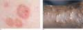
Figure 50.35
‘Cluster of jewels’ or ‘ring of pearls’ sign. The peculiar appearance of vesicles in an annular pattern or along the edge of a lesion is frequently se...
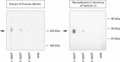
Figure 50.39
Anti‐p200 pemphigoid: serum autoantibodies against the p200 antigen and laminin γ1. By Western blotting with extract of human dermis a 200 kDa protein...
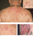
Figure 50.43
Epidermolysis bullosa acquisita, inflammatory variant. Erosions and tense blisters (insert) on the upper back (a), milia (b), and erosions of the bucc...
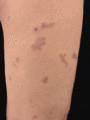
Figure 50.47
Bullous systemic lupus erythematosus. Erythematous macules and patches and a flaccid blister some days after the initiation of systemic corticosteroid...
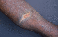
Figure 50.51
Cicatricial pemphigoid. Tense vesicles, erosions, milia and scarring on the right arm of a 24‐year‐old Ugandan man. Few oral lesions were present with...
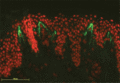
Figure 50.55
Dermatitis herpetiformis. Direct immunofluorescence demonstrating granular IgA deposition in the dermal papillae.
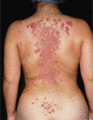
Figure 50.4
Pemphigus vulgaris. Cutaneous lesions typically affect the chest and back in addition to the scalp. (Courtesy of Dr R.J. Pye, Addenbrooke's Hospital,...
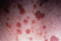
Figure 50.8
Pemphigus foliaceus. Lesions frequently have a fine superficial scale, sometimes as a collarete.
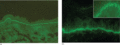
Figure 50.12
Direct immunofluorescence microscopy of bullous pemphigoid. In a perilesional biopsy, linear binding of immunoglobulin G (IgG) at the dermal–epidermal...
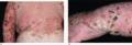
Figure 50.16
Classical bullous pemphigoid. Tense blisters, erosions, and partly haemorhagic crusts on the back and left arm (a) and left hand (b).
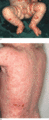
Figure 50.20
Childhood bullous pemphigoid. Disseminated tense blisters, erosions and crusts on the lower abdomen, genitalia and lower extremities in an infant (a)....
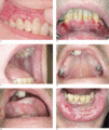
Figure 50.24
(a–f) Oral lesions in mucous membrane pemphigoid.
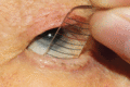
Figure 50.28
Fornix meter. Measurement of the fornix depth by an experienced ophthalmologist is important for the objective assessment of ocular disease activity. ...
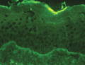
Figure 50.32
Direct immunofluorescence microscopy of a perilesional biopsy in linear IgA disease. Linear deposition of IgA at the dermal–epidermal junction.
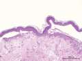
Figure 50.36
Lesional H&E histopathology of anti‐p200 pemphigoid. Subepidermal splitting and a dense neutrophilic infiltration below the blister.
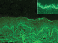
Figure 50.40
Direct immunofluorescence microscopy of a perilesional biopsy of a patient with epidermolysis bullosa acquisita. Linear deposition of IgG at the derma...
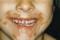
Figure 50.44
Childhood epidermolysis bullosa acquisita, inflammatory variant. Perioral yellowish crusts and erosions.
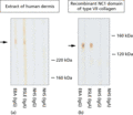
Figure 50.48
Serum autoantibodies in bullous systemic lupus erythematosus (BLSE) type I. IgA autoantibodies label the 290 kDa type VII collagen by immunoblotting w...
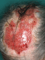
Figure 50.52
Brunsting–Perry pemphigoid. Erosions and some crusts on the scalp of a 76‐year‐old woman. By direct immunofluorescence (IF) microscopy linear deposits...
