Figure 66.1
Histology of Darier disease demonstrating: (a) acantholysis with suprabasal clefting; and (b) in close up, rounded dyskeratotic cells with eosinophili...

Figure 66.5
Nail dystrophy of Darier disease: (a) fragile nails with longitudinal splitting and terminal notching; and (b) early nail changes in a 28‐year‐old wom...

Figure 66.9
Guttate leukoderma: (a) hypopigmented macules and papules in pigmented skin (Courtesy of Dr Yoseph Legesse, University of Addis Ababa, Ethiopia); and...

Figure 66.13
Lesions of Hailey–Hailey disease: (a) typical fissured plaque in the axilla; (b) inflamed, macerated and fissured lesions of the groin in a 40‐year‐ol...

Figure 66.2
Lesions of Darier disease: (a) profuse keratotic papules in the seborrhoeic areas (b) early keratotic papules developing on the sun‐exposed skin of th...

Figure 66.6
Palmoplantar lesions of Darier disease: (a) pitting in a 15‐year‐old boy; (b) palm print demonstrating interruptions to the print pattern; and (c) ker...

Figure 66.10
Epidermal naevus following the lines of Blaschko; the clinical and histological appearances are those of Darier disease.

Figure 66.14
Extra‐epidermal lesions in patients with Hailey–Hailey disease: (a) linear white bands in the nails; terminal notches of Darier disease are not found;...

Figure 66.3
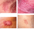
Darier's disease: (a) confluent papules forming irregular, warty, fissured plaques in the axilla; and (b) confluent inflamed and eroded lesions of the...

Figure 66.7
Oral mucosal involvement in Darier disease: (a) umbilicated and cobblestone papules in a 31‐year‐old woman (Courtesy of Dr R. I. Macleod, Royal Victo...

Figure 66.11
(a, b) Herpes simplex superinfection of skin lesions in a patient with Darier disease.

Figure 66.4
Acrokeratosis verruciformis (AKV) in Darier disease: (a) plane wart‐like lesions on the dorsal hands in a 15‐year‐old boy; (b) ‘church spire’ pattern ...

Figure 66.8
Variant forms of Darier disease: (a) cornifying plaques on the lower legs; (b) multiple comedones and deep pitted scars; and (c) haemorrhage into palm...

Figure 66.12
Lesional skin in Hailey–Hailey disease shows prominent acantholysis throughout the spinous layer, giving a so‐called ‘dilapidated brick wall’ appearan...

