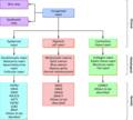
Figure 75.1
Photomicrographs of an epidermal naevus demonstrating an area of thickened epidermis with papillomatosis and mild hyperkeratosis. Original magnificati...

Figure 75.5
(a) Single sebaceous naevus on the cheek, with a yellowish hue and characteristic greasy texture. (b) Multiple pigmented sebaceous epidermal naevi on ...

Figure 75.9
Photomicrographs of a typical congenital melanocytic naevus demonstrating a predominantly dermal lesion composed of bland naevus cells that extend aro...

Figure 75.13
Benign proliferative nodule in a large congenital melanocytic naevus, present from birth and stable in behavior.

Figure 75.17
Two congenital blue naevi on the foot of a child with multiple similar lesions from birth.

Figure 75.21
Photomicrograph of aplasia cutis demonstrating an area of epidermal flattening associated with underlying fibrosis and loss of normal adnexal structur...

Figure 75.2
Photomicrograph of inflammatory linear verrucous epidermal naevus demonstrating marked epidermal thickening with hyperkeratosis and an associated infl...

Figure 75.6
Photomicrographs of an eccrine naevus demonstrating prominent eccrine glands within the dermis. Original magnifications (a) 40× and (b) 100× (H&E).

Figure 75.10
Photomicrographs demonstrating a proliferative nodule arising in a congenital melanocytic naevus composed of sheets of bland naevus cells forming a di...

Figure 75.14
Multiple neuroid‐type proliferations in a congenital melanocytic naevus.

Figure 75.18
Single naevus spilus on the face showing a café‐au‐lait macule background with superimposed darker areas.

Figure 75.22
Photomicrographs of a smooth muscle hamartoma demonstrating a poorly circumscribed dermal lesion composed of abnormal bundles of smooth muscle within ...

Figure 75.3
Photomicrographs of a typical sebaceous naevus demonstrating a lesion composed of prominent sebaceous glands within the dermis. Original magnification...

Figure 75.7
Photomicrographs of a desmoplastic trichoepithelioma arising in a sebaceous naevus demonstrating epithelial strands infiltrating the dermis surrounded...

Figure 75.11
Photomicrograph of a melanoma developing within a congenital melanocytic naevus demonstrating a proliferative subpopulation, which infiltrates widely ...

Figure 75.15
Clinical management algorithm for neurological investigation and follow‐up of patients with congenital melanocytic naevus (CMN). CNS, central nervous ...

Figure 75.19
Photomicrographs of a connective tissue naevus demonstrating a poorly circumscribed dermal lesion composed of abnormal bundles of collagen and admixed...

Figure 75.23
Phakomatosis pigmentovascularis type II (or cesioflammea type), showing the characteristic combination of (a) a capillary malformation (port‐wine stai...

Figure 75.4
Extensive inflammatory linear verrucous epidermal naevus on the lower limbs in a Blaschko‐linear distribution.

Figure 75.8
Photomicrograph of a syringocystadenoma arising in a sebaceous naevus showing a prominent papillary architecture. Original magnification 20× (H&E).

Figure 75.12
Congenital melanocytic naevi (CMN). (a) Single CMN on the face showing marked hypertrichosis. (b) Multiple CMN of different sizes on the trunk.

Figure 75.16
Photomicrographs of a blue naevus demonstrating spindle‐shaped pigmented naevus cells extending into the deep dermis. Original magnifications (a) 40× ...

Figure 75.20
Collagenoma‐type connective tissue naevus on the lower abdomen.

Figure 75.24
Unified classification of congenital naevi by clinical, histopathological and genetic criteria

