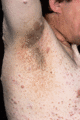
Figure 80.1
Neurofibromatosis: axillary freckling and multiple neurofibromas. (Courtesy of Professor J. Harper, Great Ormond Street Hospital, London, UK.)
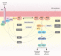
Figure 80.5
The Ras/mitogen‐activated protein kinase (MAPK) signal transduction pathway. The MAPK signalling pathway of protein kinases is critically involved in ...
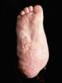
Figure 80.2
Neurofibromatosis: extensive neurofibroma of the foot. (Courtesy of Professor J. Harper, Great Ormond Street Hospital, London, UK.)
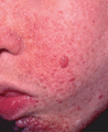
Figure 80.6
Tuberous sclerosis: angiofibromas. (Courtesy of Professor J. Harper, Great Ormond Street Hospital, London, UK.)
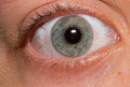
Figure 80.3
Neurofibromatosis: Lisch nodules (pigmented iris hamartomas). (Courtesy of Professor J. Harper, Great Ormond Street Hospital, London, UK.)
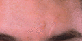
Figure 80.7
Tuberous sclerosis: fibromatous nodule on the forehead. (Courtesy of Professor J. Harper, Great Ormond Street Hospital, London, UK.)
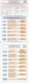
Figure 80.4
Manchester checklist for screening for neurofibromatosis. (Courtesy of Dr Sue Huson.)
