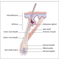
Figure 89.1
Diagram of an anagen hair follicle.
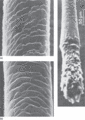
Figure 89.5
(a) Surface view of weathered cuticular scales in the distal portion of the hair shaft. (b) Surface view of undamaged cuticular scales in the proximal...
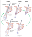
Figure 89.9
The hair cycle. (From Olsen 1994. Reproduced with permission of McGraw‐Hill.)
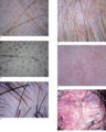
Figure 89.13
Trichoscopic images. (a) Yellow dots in alopecia areata. (b) Black dots and broken hairs in alopecia areata. (c) Variation in hair fibre diameter in f...
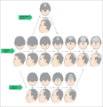
Figure 89.17
Hamilton–Norwood scale for grading male pattern hair loss (From Norwood 1975 [ ]. Reproduced with permission of Wolters Kluwer Health.)
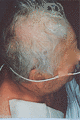
Figure 89.21
Acute telogen effluvium.
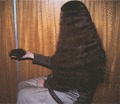
Figure 89.25
Idiopathic chronic telogen effluvium.
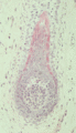
Figure 89.29
An early anagen follicle in alopecia totalis, showing vacuolation in the hair matrix epithelium around the upper pole of the dermal papilla.
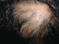
Figure 89.33
Alopecia areata showing regrowth of hair at sites of intralesional corticosteroid injection (triamcinolone acetonide).
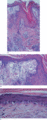
Figure 89.37
Discoid lupus erythematosus. (a) Low‐power photomicrograph showing follicular plugging, superficial and deep perivascular and periappendageal lymphocy...
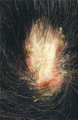
Figure 89.41
Folliculitis decalvans showing active pustulation and scarring.
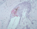
Figure 89.45
Histology of trichotillomania showing fragmentation of the hair shaft.
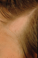
Figure 89.49
Triangular alopecia.
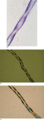
Figure 89.53
Trichorrhexis invaginata in Netherton syndrome.
Figure 89.57
Trichothiodystrophy showing alternating bright and dark zones under a polarizing microscope. (Courtesy of D. Van Neste, Brussels.)
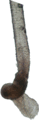
Figure 89.61
Plucked anagen hair in the loose anagen syndrome, showing a vestigial root sheath and a ruffled cuticle.
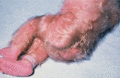
Figure 89.65
Congenital hypertrichosis lanuginosa. (Courtesy of Dr Partridge, Leamington, UK.)
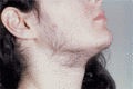
Figure 89.69
Facial hirsutism: in this case it was not associated with any systemic disease or detectable biochemical endocrine abnormality.
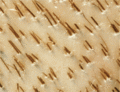
Figure 89.2
Grouping of hairs in follicular units on the human scalp. In some groups, multiple hairs emerge from a single follicular opening.
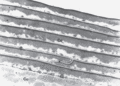
Figure 89.6
Cross‐section through hair shaft showing cuticle layers (Cu) surrounding the central cortex (Co). Transmission electron micrograph, silver methenamine...
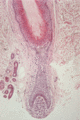
Figure 89.10
Scalp hair follicle in the anagen II stage of development. The club hair from the previous cycle is still present within the follicle. (Courtesy of D...
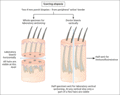
Figure 89.14
Biopsy – scarring alopecia protocol.
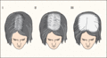
Figure 89.18
Ludwig scale for grading female pattern hair loss. (From Ludwig 1977 [ ]. Reproduced with permission of John Wiley.)

Figure 89.22
Diffuse alopecia in association with hypothyroidism.
Figure 89.26
‘Porrigo decalvans’ (alopecia areata). Plate XL from Thomas Bateman's atlas Practical Synopsis of Cutaneous Diseases , 1819.
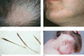
Figure 89.30
Alopecia areata. (a) Patch of alopecia areata showing broken ‘exclamation mark hairs’ towards the margins. (b) Close‐up of exclamation mark hairs. (c)...
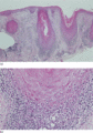
Figure 89.34
Lichen planopilaris. (a) Low‐power photomicrograph showing follicular plugging and a periappendageal inflammatory infiltrate. (b) Base of the hair fol...
Figure 89.38
Positive linear immunofluorescence to IgG: the lupus band test. (Courtesy of Dr G. Mason, Melbourne, Australia.)
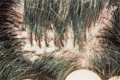
Figure 89.42
Tufted folliculitis.
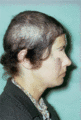
Figure 89.46
Trichotillomania showing a characteristic ‘tonsure’ pattern with the scalp margin hair spared.
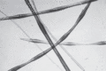
Figure 89.50
Monilethrix with swollen (node) and narrow (internode) fluctuations in the hair bore.
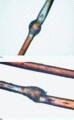
Figure 89.54
Netherton syndrome. (a) An invaginate node showing partial twisting of the hair at the upper pole. (b) An invaginate node acting as a point of weaknes...
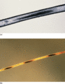
Figure 89.58
Pili annulati. (a) Hair shaft by transmitted light showing an abnormal dark band (central part) caused by multiple cortical air spaces. This correspon...
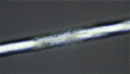
Figure 89.62
Focal loss of the cuticle in a weathered hair.
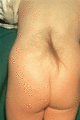
Figure 89.66
Lumbosacral hypertrichosis (‘faun tail’), here associated with diastematomyelia.
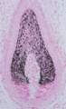
Figure 89.70
Human anagen follicle, showing pigment donation to the hair cortex. There is pigment incontinence in the dermal papilla. Masson–Fontana stain.
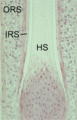
Figure 89.3
Longitudinal section through the suprabulbar region of an anagen follicle showing the keratogenous region of the hair shaft (HS). The inner root sheat...
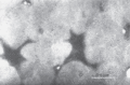
Figure 89.7
Cross‐section of transformed cortical cells of human hair. The relatively translucent filaments, set in a more dense sulphur‐rich matrix, appear as co...
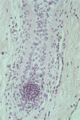
Figure 89.11
Human hair follicle in mid‐catagen. There is a prominent glassy membrane surrounding the regressing epithelial column. The dermal papilla has rounded ...
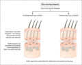
Figure 89.15
Biopsy – non‐scarring alopecia protocol.
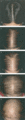
Figure 89.19
Pattern of frontal hair loss in women with androgenetic alopecia. Stage 1 is normal.
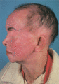
Figure 89.23
Acquired zinc deficiency resulting from prolonged parenteral feeding and inadequate zinc supplementation.
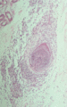
Figure 89.27
Lymphocytic inflammatory infiltrate surrounding an anagen hair bulb in alopecia areata.
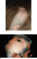
Figure 89.31
(a) Sparing of white hairs in a patch of alopecia areata.(b) Regrowth of hypopigmented hair in alopecia areata.
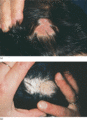
Figure 89.35
(a) Scarring alopecia caused by lichen planus showing active lesions.(b) More advanced lichen planus showing follicular plugs and scarring.
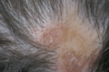
Figure 89.39
Discoid lupus erythematosus showing scarring alopecia with scalp erythema and follicular plugging.
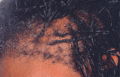
Figure 89.43
Traction alopecia from braiding.
Figure 89.47
Trichodysplasia spinulosa.
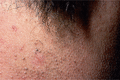
Figure 89.51
Monilethrix on the nape of the neck showing follicular keratoses and short, broken hairs.
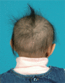
Figure 89.55
Localized autosomal recessive hypotrichosis due to a dsg4 mutation.
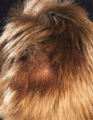
Figure 89.59
Woolly hair naevus.
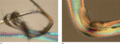
Figure 89.63
(a) Knotting of single and multiple hairs contributes to hair shaft trauma. (b) Braiding damages the hair shaft cuticle.
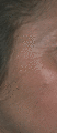
Figure 89.67
Facial hypertrichosis in porphyria cutanea tarda.
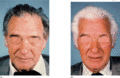
Figure 89.71
Rapid greying of the hair caused by alopecia areata. (a) A patient with slight greying of the hair, and (b) the same patient shown 1 week later. (Cou...
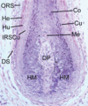
Figure 89.4
Anagen hair bulb. Co, hair cortex; Cu, hair cuticle; DP, dermal papilla; DS, dermal sheath; He, Henle layer; HM, hair matrix; Hu, Huxley layer; IRSCu,...
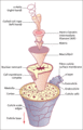
Figure 89.8
Exploded view of the major structural components comprising a human hair fibre. Pigment granules that are normally dispersed throughout the cortex are...
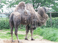
Figure 89.12
Bactrian camel in a spring moult.
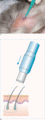
Figure 89.16
(a, b) Orientation of the punch biopsy in the direction of hair growth.
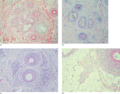
Figure 89.20
Histopathology of female pattern hair loss. (a) Increase in telogen forms – three of five follicles in a follicular unit are in telogen. (b) Follicula...
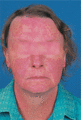
Figure 89.24
Hair loss and photosensitivity caused by systemic lupus erythematosus.
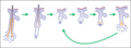
Figure 89.28
Proposed pathodynamic changes in alopecia areata. An inflammatory attack on anagen follicles precipitates follicles into telogen. Follicles re‐enter a...
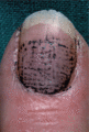
Figure 89.32
An organized pattern of pitting present on all fingernails 8 months prior to the onset of alopecia areata. The pits are highlighted with mascara.
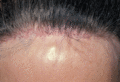
Figure 89.36
Frontal fibrosing alopecia showing scarring alopecia affecting the frontal hairline with follicular erythema and scale.
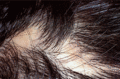
Figure 89.40
Pseudopelade of Brocq.
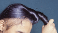
Figure 89.44
Traction alopecia in a Sikh boy.
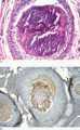
Figure 89.48
Trichodysplasia spinulosa histopathology. (a) Aberrant keratinization of the inner root sheath. (b) Positive immunohistochemical staining for polyomav...
Figure 89.52
Pili torti. (a) Light micrograph showing 180° twists. (b) A 6‐month‐old boy with Menkes syndrome. (c) A 27‐year‐old woman with no personal or family h...
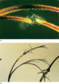
Figure 89.56
Trichorrhexis nodosa. (a) Polarized light examination demonstrates the splayed cortical fibres radiating from the transverse fracture in a trichorrhex...
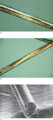
Figure 89.60
Uncombable hair syndrome. (a) The triangular cross‐section of the hair contributes to its stiffness. (b) Light microscopy revealing grooving when usin...
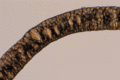
Figure 89.64
Appearance of normal scalp hair after exposure to a naked flame. Bubbles form within the cortex.
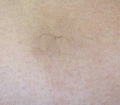
Figure 89.68
Localized hypertrichosis at the site of underlying panniculitis.
