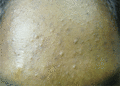
Figure 90.1
Predominantly comedonal acne. (Courtesy of Dr S. Chow, KL Skin Centre, Malaysia.)
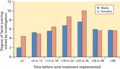
Figure 90.5
There is a correlation between acne scarring and duration of acne. Scarring is more likely to occur with delays in treatment. (Adapted from Layton et...
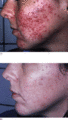
Figure 90.9
(a) Non‐classical congenital adrenal hyperplasia in a 16 year old with oligomenorrhoea pretreatment. (b) Post‐therapy with 2 mg oral prednisolone dail...
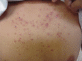
Figure 90.13
Monomorphic inflammatory papules and pustules associated with corticosteroid use. (Courtesy of Dr S. Chow, KL Skin Centre, Malaysia.)
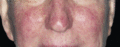
Figure 90.17
Epidermal growth factor receptor inhibitor producing follicular acneform eruption on the face of a patient receiving treatment for colonic cancer.
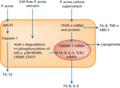
Figure 90.21
Effects of P. acnes extracts on human sebocytes. CREBP, cAMP response element‐binding protein; hBD‐2, human β‐defensin 2; IL, interleukin; p38‐MAPK,...
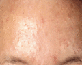
Figure 90.25
Comedonal acne with mid‐facial distribution. This distribution is associated with poor prognosis.
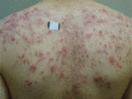
Figure 90.29
Severe acne of the back with many inflammatory papules and pustules.
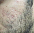
Figure 90.33
Atrophic scarring with associated inflammatory change. (Courtesy of J. Del Rosso Las Vegas Skin & Cancer Clinic, Las Vegas, Nevada, USA.)
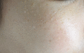
Figure 90.37
Multiple milia on the upper cheek. (Courtesy of Dr J. Del Rosso, Las Vegas Skin & Cancer Clinic, Las Vegas, Nevada, USA.)
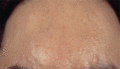
Figure 90.41
Sebaceous gland hyperplasia on the forehead. (From Zouboulis CC, Boschnakow A. Chronological ageing and photoageing of the human sebaceous gland. Clin...
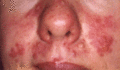
Figure 90.45
Rosacea on the mid‐face with periorbital sparing.
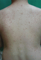
Figure 90.49
Pityrosporum folliculitis in an immunocompromised male. (Courtesy of Dr S. Chow, KL Skin Centre, Malaysia.)
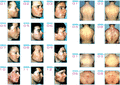
Figure 90.53
The Leeds photometric acne grading scale used to assess the face and trunk. (Reproduced from O'Brien et al . 1998 [ ] with permission from the Leeds...
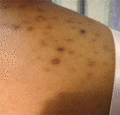
Figure 90.57
Post‐inflammatory hyperpigmentation in Fitzpatrick type IV skin.
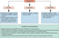
Figure 90.61
Treatment algorithm for severe acne. BPO, benzoyl peroxide.
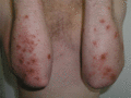
Figure 90.65
Staphylococcus aureus colonizing discoid eczema induced by oral retinoids.
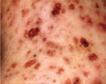
Figure 90.69
Erosive crusting lesions on the back of a young male with acne fulminans.
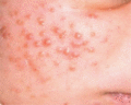
Figure 90.73
Neonatal cephalic pustulosis.
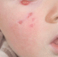
Figure 90.77
Infantile acne may involve cystic lesions and scarring. (Courtesy of Dr J. Ravenscroft, Queens Medical Centre, University of Nottingham, UK.)
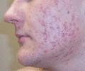
Figure 90.2
Moderate to severe inflammatory acne including a mixture of non‐inflammatory and inflammatory lesions with seborrhoea.
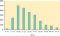
Figure 90.6
The age distribution of acne is widening in both sexes.
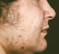
Figure 90.10
Seborrhoea, acne, hirsutism and/or androgenic alopecia (SAHA) syndrome.
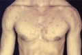
Figure 90.14
Severe acne vulgaris in a male body builder.
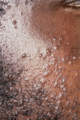
Figure 90.18
Pomade acne.
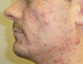
Figure 90.22
Moderate to severe inflammatory acne on the face.

Figure 90.26
Sandpaper comedones on the forehead.
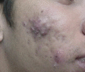
Figure 90.30
Nodular acne of the right cheek with scars. (Courtesy of Dr S. Chow, KL Skin Centre, Malaysia.)
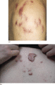
Figure 90.34
(a) Hypertrophic scarring of the shoulders in the context of moderate to severe acne. (b) Keloid scarring on the trunk associated with mild acne. (Co...
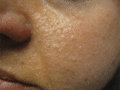
Figure 90.38
Fibrofolliculomas: Birt–Hogg–Dube syndrome. (Courtesy of Dr J. Del Rosso, Las Vegas Skin & Cancer Clinic, Las Vegas, Nevada, USA.)
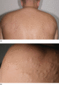
Figure 90.42
(a) Steatocystoma multiplex of the back. (b) Steatocystoma close‐up of the multiple cystic lesions. (Courtesy of Dr N. Veien, the Dermatology Clinic,...
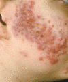
Figure 90.46
Rosacea fulminans (synonymous with pyoderma faciale).

Figure 90.50
Folliculitis keloidalis affecting the nape of the neck and hairline resulting in cicatricial alopecia.
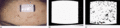
Figure 90.54
Sebutape analysis. Comparison of two Sebutapes demonstrating the difference between patients with and without acne. (a) Sebutape assessment of sebum. ...
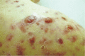
Figure 90.58
Pyogenic granulomas in severe acne.
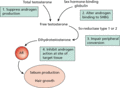
Figure 90.62
Potential mechanism of action(s) using antiandrogens in the management of acne. AR, androgen receptor; SHBG, sex hormone binding globulin.
Figure 90.66
Hyfrecation of macrocomedones.
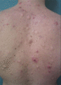
Figure 90.70
Acne conglobata of the back with multiple inflammatory lesions, grouped comedones, cysts and scarring.
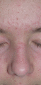
Figure 90.74
Mid‐facial comedones are associated with poor prognosis.
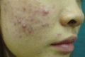
Figure 90.3
Post‐inflammatory macules and pigment changes interspersed with inflammatory acne. (Courtesy of Dr S. Chow, KL Skin Centre, Malaysia.)
Figure 90.7
(a) Acne in an adult female with polycystic ovary syndrome associated with hirsutism and seborrhoea. (b) Ultrasound scan showing cysts (+) on the ovar...
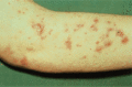
Figure 90.11
The unusual extent of acne in a patient with Apert syndrome.
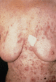
Figure 90.15
Lithium‐induced acne. (Courtesy of Dr V. M. Yates, Royal Bolton Hospital, UK.)
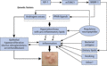
Figure 90.19
Inflammatory cascades involved in acne pathogenesis. SREBP‐1, sterol response element‐binding protein 1. (Modified from Zouboulis et al. 2005 [ ].)
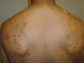
Figure 90.23
Acne on the back showing sparing of the central back.
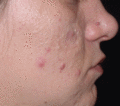
Figure 90.27
Multiple macrocomedones interspersed with some inflammatory lesions on the cheeks of a female patient with acne. (Courtesy of Professor M. Jackson, U...
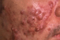
Figure 90.31
Nodular/conglobate acne with sinus tracts. (Courtesy of Dr C. L. Goh, National Skin Centre, Singapore.)

Figure 90.35
Acné excoriée on the forehead of a female. (Courtesy of Dr C. L. Goh, National Skin Centre, Singapore.)
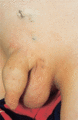
Figure 90.39
A patient with comedo naevus (naevus comedonicus) predominantly consisting of blackheads on the lower abdomen.
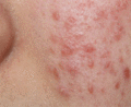
Figure 90.43
Granulomatous rosacea synonymous with acne agminata seen on the cheek.

Figure 90.47
Perioral dermatitis demonstrating small papules on an erythematous base.
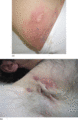
Figure 90.51
(a) Hidradenitis suppurativa of the groin showing inflammation, comedonal lesions and cribriform scarring. (b) Hidradenitis suppurativa of the right a...
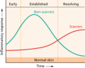
Figure 90.55
Immune responses vary between scarring and non‐scarring acne patients.
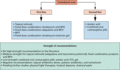
Figure 90.59
Treatment algorithm for comedonal acne. BPO, benzoyl peroxide.
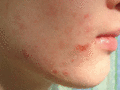
Figure 90.63
Retinoid dermatitis as a result of oral retinoids.
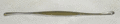
Figure 90.67
Comedo extractor.
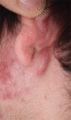
Figure 90.71
Patients with acne conglobata present with grouped comedones and deep‐seated inflammatory lesions.
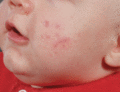
Figure 90.75
Infantile acne on the cheek. (Courtesy of Dr J. Ravenscroft, Queens Medical Centre, Nottingham, UK.)
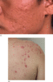
Figure 90.4
Acne scarring. (a) Atrophic scarring on the cheeks. (Courtesy of Dr C. L. Goh, National Skin Centre, Singapore.) (b) Hypertrophic keloid scarring of ...
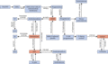
Figure 90.8
Schematic representation of the role of 21‐hydroxylase in the adrenal steroid genesis pathway. See text for abbreviations.
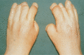
Figure 90.12
The typical appearance of the fingers of a patient with Apert syndrome.
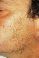
Figure 90.16
Chloracne – multiple comedonal lesions on the face.

Figure 90.20
Neuropeptide–cytokine/chemokine signalling in human sebaceous glands and human sebocytes. Red: promoter of inflammation. Green: inhibition of inflamma...
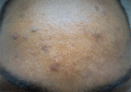
Figure 90.24
Post‐inflammatory erythema and pigment changes on the forehead. (Courtesy of Dr J. Del Rosso, Las Vegas Skin & Cancer Clinic, Las Vegas, Nevada, USA....
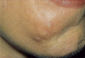
Figure 90.28
Submarine comedones. This patient required stretching of the skin in order for them to be seen.
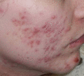
Figure 90.32
Inflammatory macules contribute to the erythema seen in acne.
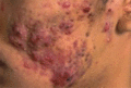
Figure 90.36
Granulomatous acne of the face. (Courtesy of Dr C. L. Goh, National Skin Centre, Singapore.)
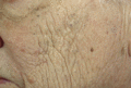
Figure 90.40
Favre–Racouchot syndrome (senile comedones). (Courtesy of Dr J. Del Rosso, Las Vegas Skin & Cancer Clinic, Las Vegas, Nevada, USA.)
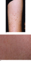
Figure 90.44
(a) Keratosis pilaris of the upper arms associated with some inflammation and excoriation. (b) A close‐up view of keratosis pilaris.
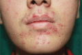
Figure 90.48
Gram‐negative folliculitis after long‐term antibiotic use showing multiple pustules. (Courtesy of Dr S. Chow, KL Skin Centre, Malaysia.)
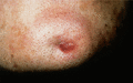
Figure 90.52
Dental sinus confused with persistent facial acne nodule.
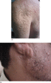
Figure 90.56
(a) Severe scarring of the arms and back showing soft distensible scars as a result of acne. (Courtesy of Dr S. Chow, KL Skin Centre, Malaysia.) (b) ...
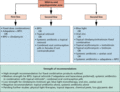
Figure 90.60
Treatment algorithm for mild to moderate inflammatory acne. BPO, benzoyl peroxide.
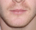
Figure 90.64
Cheilitis and fissure of the lower lip induced by oral retinoids.
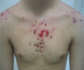
Figure 90.68
Acne fulminans in a young male.
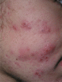
Figure 90.72
Patient with acne conglobata present with abscesses and cysts, causing interconnecting sinus tracts.
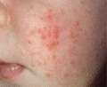
Figure 90.76
Neonatal acne presenting in the first few weeks of life.
