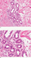
Figure 94.1
(a) A normal eccrine unit composed of secretory glands and ducts. Magnification 10× (H&E). (b) Closer view of eccrine glands showing the double layer ...
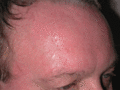
Figure 94.5
Cranio‐facial hyperhidrosis. It may be sufficiently profuse to drip off the face and wet the hair.
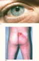
Figure 94.9
Ross syndrome. (a) The pupils are tonic, asymmetrical and irregular in outline. (b) Most of the skin is anhidrotic but the remaining areas of enervate...
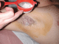
Figure 94.2
Identifying the extent of axillary hyperhidrosis – the skin has been cleaned with povidobe iodine solution and then sprinkled with corn starch powder ...
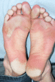
Figure 94.6
94.6Plantar hyperhidrosis showing maceration of the plantar keratin and secondary pitted keratolysis due to infection with Kytococcus sedentarius .
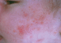
Figure 94.10
Miliaria rubra affecting the cheeks of an infant. (Courtesy of Dr Richard Logan, Bridgend, UK.)
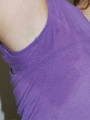
Figure 94.3
Axillary hyperhidrosis: patients often wear white or black garments as the wetness is not as visibly obvious as with coloured clothes.
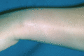
Figure 94.7
Circumscribed (naevoid) hyperhidrosis on the wrist. There is a solitary area of hyperhidrosis with normal sweating elsewhere on the rest of the skin.
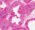
Figure 94.11
Normal apocrine glands lined by cells with abundant eosinophilic cytoplasm and decapitation secretions. Magnification 40× (H&E). (Courtesy of Dr Arti...
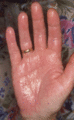
Figure 94.4
Palmar hyperhidrosis.
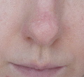
Figure 94.8
Granulosis rubra nasi in young adult female showing localised hyperhidrosis with beads of sweat on nose and philtrum together with multiple vesicles a...
Figure 94.12
Axillary apocrine miliaria (Fox–Fordyce disease).
