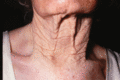
Figure 96.1
Actinic elastosis on the neck of an elderly female patient.
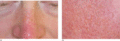
Figure 96.5
(a,b) Adult colloid milium with multiple tiny yellowish translucent papules on the dorsum of the nose (a) with close‐up view of papules on the cheek (...
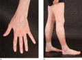
Figure 96.9
(a,b) Severe generalized cutaneous atrophy in a 29‐year‐old female as the result of using inhaled corticosteroids for asthma since the age of 7; (b) n...
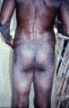
Figure 96.13
Atrophy due to onchocerciasis. (From Murdoch et al . 1993 [ ], courtesy of Dr M. Murdoch, West Hertfordshire Hospitals NHS Trust, Hertfordshire, UK....
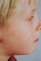
Figure 96.17
Spontaneous atrophic scarring of the cheeks (varioliform atrophy).
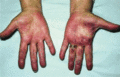
Figure 96.21
Paroxysmal haematoma of the finger. (Courtesy of Dr J. Verbov, Royal Liverpool University Hospitals, Liverpool, UK.)
Figure 96.25
Secondary anetoderma in chickenpox scar. (From Veraldi A, Schianchi R, Chickenpox, impetigo, and anetoderma Pediatric Dermatology 2006;23:305–6. Wit...
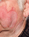
Figure 96.29
Typical actinic granulomas on the face and neck of an elderly man.
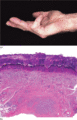
Figure 96.33
(a,b) Palmar fascial fibromatosis: clinical image illustrating (a) typical fixed contraction of the little finger, and (b) low‐power photomicrograph s...
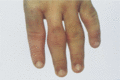
Figure 96.37
Pachydermodactyly. (Courtesy of Dr A. Chamberlain, Churchill Hospital, Oxford, UK.)
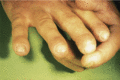
Figure 96.41
Vinyl chloride‐induced osteolysis affecting fingertips.
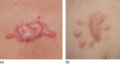
Figure 96.45
(a,b) Contrast between two scars from the presternal area: (a) spontaneous keloid, and (b) hypertrophic scar following excision of benign mole; the fo...
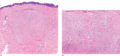
Figure 96.49
(a,b) Keloid nodule: large well‐circumscribed dermal nodule sparing papillary dermis (a); higher‐power view with haphazardly arranged thick sclerotic ...
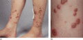
Figure 96.53
(a,b) Acquired perforating dermatosis: a 48‐year‐old woman with a 25‐year history of type 1 diabetes with retinopathy and renal failure; and a 12‐year...
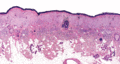
Figure 96.2
Actinic elastosis showing confluent masses of amorphous basophilic material in the papillary and upper reticular dermis with atrophy of the overlying ...
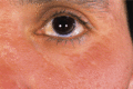
Figure 96.6
More advanced adult colloid milium manifesting as confluent plaques of the infraorbital region but with individual papules discernible at the margins....
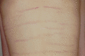
Figure 96.10
Pubertal growth striae across the back of an adolescent boy: note that these are normally all horizontally arranged right across the back (compare wit...
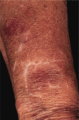
Figure 96.14
Stellate pseudoscars on the forearm of an elderly woman. There was no history of trauma.
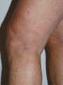
Figure 96.18
Acrodermatitis chronica atrophicans: image captured soon after commencement of antibiotic therapy; note atrophic wrinkled appearance of the skin at th...
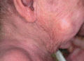
Figure 96.22
Acquired cutis laxa following a generalized inflammatory dermatitis in an 18‐month‐old child. (From Haider et al . [ ], with permission from John Wil...
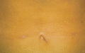
Figure 96.26
Idiopathic mid‐dermal elastolysis. (Courtesy of Dr L. Ostlere, St George's Hospital, London, UK.)
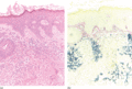
Figure 96.30
(a,b) Annular elastolytic giant cell granuloma: low‐power view showing intense granulomatous inflammation (a) and elastorrhexis with loss of elastic f...
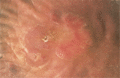
Figure 96.34
Plantar fibromatosis.
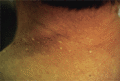
Figure 96.38
White fibrous papulosis of the neck. (Courtesy of Professor H. Shimizu, Sapporo Hospital, Tokyo, Japan.)
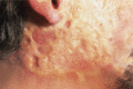
Figure 96.42
Scleroderma and scarring of the face due to porphyria cutanea tarda.
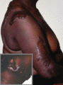
Figure 96.46
Extensive disfiguring keloids affecting an Afro‐Caribbean woman.
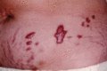
Figure 96.50
Fresh keloids arising in striae gravidarum 3 years after pregnancy.
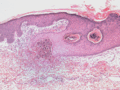
Figure 96.54
Elastosis perforans serpiginosa: note acanthotic epidermis growing downward in order to surround and engulf a focus of basophilic elastotic debris. (...

Figure 96.3
(a,b) Nodular actinic elastosis with comedones and cysts (Favre–Racouchot syndrome): early stages in a 78‐year‐old woman (a) and advanced stage in an ...
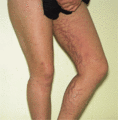
Figure 96.7
Striae of the legs due to long‐term application of a potent topical steroid in a young woman with psoriasis.
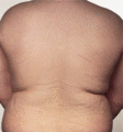
Figure 96.11
Striae due to obesity in a young man.
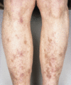
Figure 96.15
Brown pseudoscars of the legs due to diabetic dermopathy. There was no history of trauma.
Figure 96.19
Follicular atrophoderma in Conradi syndrome.
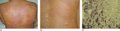
Figure 96.23
Post‐inflammatory elastolysis and cutis laxa (Marshall syndrome) in a 6‐year‐old boy showing acute inflammatory phase (a) progressing to large plaques...
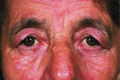
Figure 96.27
Blepharochalasis.
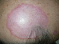
Figure 96.31
Actinic granuloma on a bald scalp. (Courtesy of Professor Luis Requena, Universidad Autónoma de Madrid, Spain.)
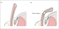
Figure 96.35
(a) Normal erect penis. (b) Erect penis with deformation from Peyronie disease showing a fibrous plaque causing ‘waisting’.

Figure 96.39
Camptodactyly in the ring and little fingers. (From Almeida SF, Monteiro AV, Lanes RCdS. Evaluation of treatment for camptodactyly: retrospective anal...
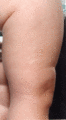
Figure 96.43
Constricting band across the thigh of a 6‐month‐old infant.
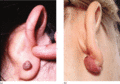
Figure 96.47
(a,b) Earlobe keloid.
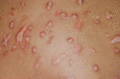
Figure 96.51
Acquired perforating dermatosis: close‐up view of the back of a 65‐year‐old woman with longstanding diabetes.
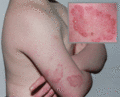
Figure 96.55
Elastosis perforans serpiginosa in a boy with Down syndrome.
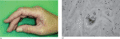
Figure 96.4
(a,b) Collagenous and elastotic marginal plaques of the hands: linear plaque involving radial aspect of the right index finger of a 49‐year‐old woman ...
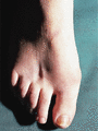
Figure 96.8
Localized atrophy due to injection of a steroid (triamcinolone 40 mg/mL) into the skin between the second and third metatarsals.
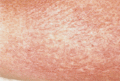
Figure 96.12
Poikilodermatous mycosis fungoides.
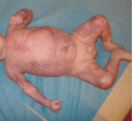
Figure 96.16
Congenital erosive and vesicular dermatosis with reticulate scarring. (From De Lange et al . 2009 [ ], with permission from John Wiley.)
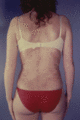
Figure 96.20
Atrophoderma of Pasini and Pierini.
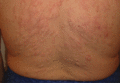
Figure 96.24
Primary anetoderma associated with antiphospholipid antibodies. (From Eungdamrong et al . [ ], with permission from Dermatology Online Journal .)
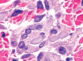
Figure 96.28
Annular elastolytic giant cell granuloma: high‐power view showing fragments of degenerate elastic fibres engulfed by multinucleate giant cells. (Cour...
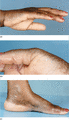
Figure 96.32
(a–c) Acrokeratoelastoidosis.
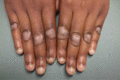
Figure 96.36
Knuckle pads. (From Hyman and Cohen [ ], with permission from Dermatology Online Journal .)
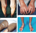
Figure 96.40
Nephrogenic systemic fibrosis: Deep involvement where fibrosis pulls down a linear band of skin on the thighs (a); tightness and hardness of the hands...
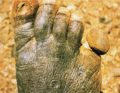
Figure 96.44
Ainhum, just before shedding of the fifth digit. (Courtesy of Dr D. Burley, Princess Margaret Hospital, Swindon, UK.)

Figure 96.48
Keloid nodules secondary to acne.
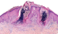
Figure 96.52
Acquired perforating dermatosis: invaginations of the epidermis enable columns of necrotic inflammatory debris to be extruded from the dermis. (Court...
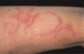
Figure 96.56
Elastosis perforans serpiginosa in a patient with vascular Ehlers–Danlos syndrome.
